List of abbreviations
Vocabulary
of micros-
copic
anatomy
specialist terms
explained in
English +
German

Every attempt was made to provide correct information and labelling, however any liability for eventual errors or incompleteness is rejected!

dieser Seite

Editor:
Dr. med.
H. Jastrow


Conditions
of use
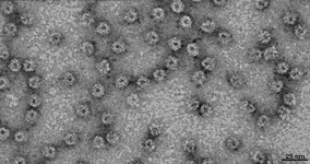
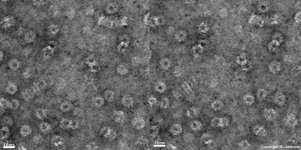
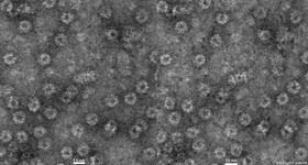
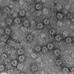
Overview proteasomes (Proteasoma):
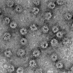
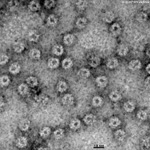
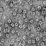
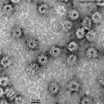
The images derive from a 20s proteasome of an egg cell from Xenopus laevis.
stereo image 1
stereo image 2
longitudinal + cross-section
Proteasomes (Terminologia histologica: Proteasoma) are large protein complexes with a hollow cylindrical shape. Other regulatrory proteins can be located at both ends of proteasome complex, whereas the protein cleavage site is located in the central channel. There is still discussion as to whether proteasomes are in reality cell organelles or just large proteins; most favour the latter interpretation. TEM images of proteasome have been produced from many different cell types. 3D image reconstructions reveal the molecular detail, with an
average length of ~ 45 nm and diameter of ~13 nm. A long channel with a diameter of ~ 6nm (resembling a pore when seen from above) is located in the centre of the cylindrial molecule. Higher resolution X-ray structural detail is also avialable. Proteasomes are present in both the cytoplasm and nucleus of all eukaryotic cells, and in the Archaeobacteria. Proteasomes play an important role in the following processes: cellular differenciation, signal transduction, regulation of metabolic activity and cell division (mitosis) by selective degradation of regulating and abnormal proteins. Functionally proteasomes are proteinases, i.e. proteins that serve for degradation of other proteins. Thereby specific enzymes link NH2-groups to proteins that are destined to be destroyed in an energy-dependent manner and link the marked proteins to several ubiquitin molecules. These ubiquitin units draw the marked protein into the channel of a proteasome, where it is split into peptides, while the linked ubiquitin molecules are recycled.

--> nucleus, cytoplasm, lysosomes
--> Electron microscopic atlas Overview
--> Homepage of the workshop


I am very grateful to Prof. Dr. rer. nat. R. Harris for the specimens. Images, page & copyright H. Jastrow.